




The biology of ageing

The twelve hallmarks
of ageing

Ageing is driven by twelve distinct but interconnected biological mechanisms. Each operates at the cellular level, each accelerates the others, and each defines a target for lifestyle and clinical intervention.

López-Otín, Blasco, Partridge, Serrano & Kroemer · Cell, 2023

01 The map

Twelve mechanisms across three connected tiers

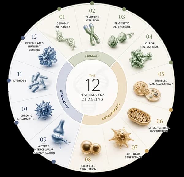
The hallmarks group into three tiers: primary causes, antagonistic responses, and integrative consequences. They do not act in isolation — each mechanism accelerates the others across the full cycle of ageing.

The 12 Hallmarks of Ageing diagram

Twelve mechanisms, one integrated system.

PrimaryCauses · 01–04Primary sources of molecular and cellular damage that accumulate throughout life.
AntagonisticResponses · 05–08Compensatory responses that become harmful when chronic or excessive.
IntegrativeConsequences · 09–12Downstream consequences that arise when damage exceeds the capacity for repair.
02 The cascade

The relationship between the three tiers

Primary hallmarks initiate damage. Antagonistic hallmarks arise as compensatory responses, then contribute to the damage themselves. Integrative hallmarks emerge when the accumulated burden exceeds the body's capacity to repair and maintain tissue.

Primary
Causes
Sources of damage present throughout life.
  • 01Genomic Instability
  • 02Telomere Attrition
  • 03Epigenetic Alterations
  • 04Loss of Proteostasis
Antagonistic
Responses
Compensatory responses that become harmful.
  • 05Disabled Macroautophagy
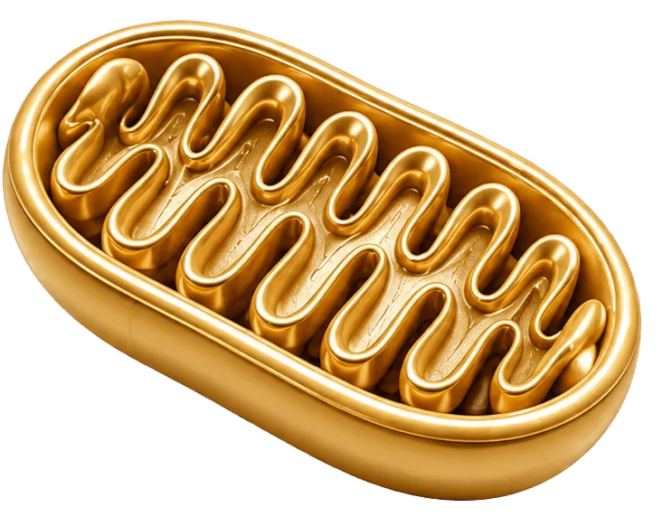
  • 06Mitochondrial Dysfunction
  • 07Cellular Senescence
  • 08Stem Cell Exhaustion
Integrative
Consequences
Consequences of unrepaired damage.
  • 09Altered Intercellular Communication
  • 10Chronic Inflammation
  • 11Dysbiosis
  • 12Deregulated Nutrient Sensing
03 The twelve

The twelve hallmarks in detail

PrimaryThe damage that initiates ageing01–04
Primary · 01–04
01

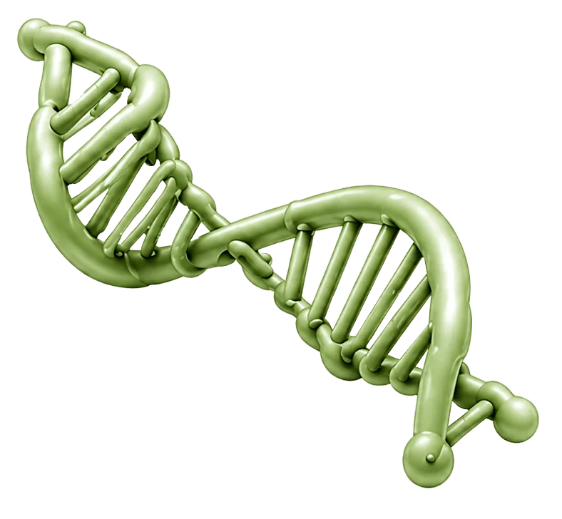
Genomic Instability

DNA Damage · cGAS-STING Signalling

Somatic DNA damage accumulates as repair capacity declines with age.

Cellular DNA is continuously damaged by radiation, chemical exposure, metabolic byproducts and replication errors. Repair systems correct most of this damage in young cells. With age, repair becomes less efficient and lesions accumulate, corrupting the information cells depend on.

Mechanism

A less recognised consequence is the mislocalisation of DNA within the cell. Chromosomal fragments form cytoplasmic micronuclei, and LINE-1 retrotransposons, normally epigenetically silenced, become derepressed and generate cytoplasmic DNA copies. The cytosolic DNA sensor cGAS, and its effector STING, detect this displaced self-DNA and activate a type I interferon response, producing sterile chronic inflammation.

Evidence

The cGAS-STING pathway is now considered a principal link between genomic damage and the chronic systemic inflammation of ageing, and is under investigation as a therapeutic target.1, 7

02

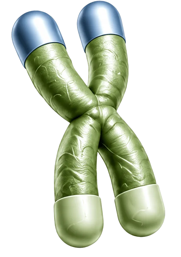
Telomere Attrition

Telomere Biology · Mitochondrial Signalling

Progressive telomere shortening limits replicative capacity and signals to cellular metabolism.

Telomeres are repetitive DNA sequences that protect chromosome ends. They shorten with each cell division until the cell can no longer divide and enters arrest or senescence.

Mechanism

Telomere function extends beyond replicative counting. When telomeres lose the protective shelterin complex, they activate the p53 pathway, which represses PGC-1α, a principal regulator of mitochondrial biogenesis. Telomere dysfunction therefore reduces mitochondrial capacity, connecting telomere maintenance directly to cellular energy metabolism.

Evidence

In a prospective study of 35 healthy older adults, a protocol of 60 daily hyperbaric oxygen sessions over 90 days increased telomere length in isolated immune cells by 20% to 38% and reduced the proportion of senescent T cells by up to 37%. The trial was small and uncontrolled, but the reported effect on telomere length exceeds that of most non-genetic interventions.3

03

Epigenetic Alterations

Epigenetics · Biological Age

Age-related changes in gene regulation, rather than gene sequence, alter cellular function.

The epigenome, comprising DNA methylation and histone modifications, determines which genes each cell expresses without changing the DNA sequence. With age this regulatory layer loses precision, and cells increasingly express genes inappropriate to their type.

Mechanism

The Information Theory of Ageing proposes that ageing results from progressive loss of epigenetic information. When cells repair DNA double-strand breaks, chromatin-modifying proteins relocate from their normal positions to sites of damage and do not fully return. Repeated over a lifetime, this relocalisation erodes the epigenetic patterns that maintain cell identity.

Evidence

In 2023, researchers engineered mice, termed ICE mice, to accumulate epigenetic disruption without added mutation, and these animals developed features of ageing. Partial reprogramming with Oct4, Sox2 and Klf4 reversed several of these features and reduced epigenetic age by up to 57%. Methylation-based epigenetic clocks now estimate biological age from the same marks.2

04

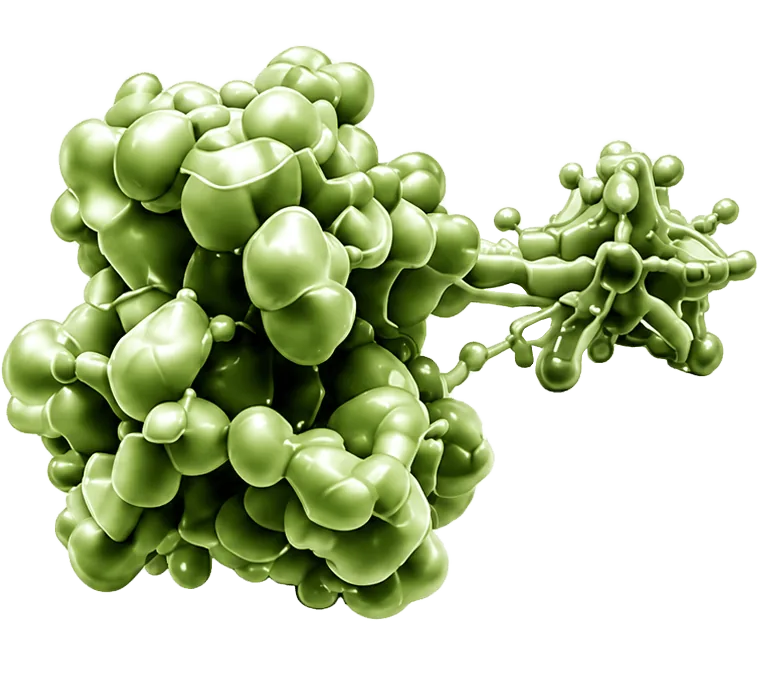
Loss of Proteostasis

Protein Homeostasis

The systems maintaining correct protein folding and turnover become less effective.

Proteins must fold into specific structures to function. Cells maintain continuous quality control to refold or degrade defective proteins. When this control declines, misfolded proteins aggregate. Protein aggregation is a common feature of Alzheimer's disease, Parkinson's disease and type 2 diabetes.

Mechanism

Two mechanisms decline in parallel. The heat-shock response, regulated by the transcription factor HSF1, produces fewer chaperones to refold stressed proteins. In addition, chaperone-mediated autophagy, which delivers damaged proteins for degradation through the LAMP2A receptor, becomes less active.

Evidence

In aged mice, restoring expression of the LAMP2A receptor improved liver function, indicating that the degradation machinery itself is a viable target, not only the aggregated proteins.1

AntagonisticCompensatory responses that become harmful05–08
Antagonistic · 05–08
05

Disabled MacroautophagyNew · 2023

Autophagy · Mitophagy

Autophagy, the cellular degradation and recycling process, declines with age.

Autophagy degrades and recycles damaged organelles, protein aggregates and other cellular components. Its activity decreases with age, allowing dysfunctional material to accumulate.

Mechanism

Macroautophagy was classified as a separate hallmark in 2023 because its specialised branches decline independently. Mitophagy, the selective removal of damaged mitochondria marked by the PINK1/Parkin system, is particularly important. Expression of TFEB, the transcription factor that coordinates the autophagy programme, also decreases with age.

Evidence

Urolithin A, a metabolite produced by gut bacteria from ellagitannins in pomegranate and walnuts, activates mitophagy. In randomised trials in middle-aged and older adults, supplementation improved muscle strength by approximately 12% and reduced C-reactive protein. A substantial proportion of people cannot produce urolithin A from diet, which links this hallmark to the composition of the gut microbiome.4

06

Mitochondrial Dysfunction

Bioenergetics · NAD+

Mitochondrial efficiency declines, affecting both energy production and cellular signalling.

Mitochondria produce most cellular energy. With age they become less efficient and generate more reactive oxygen species, which further damage mitochondrial components and reduce output.

Mechanism

Two less familiar changes contribute. Mitochondria secrete signalling molecules known as mitochondrial-derived peptides, including MOTS-c and humanin, which regulate systemic metabolism and decline with age. In parallel, levels of the coenzyme NAD+, required for both energy metabolism and the sirtuin repair enzymes, fall progressively.

Evidence

In animal studies MOTS-c improves metabolic resilience and reproduces several effects of exercise. NAD+ precursors, including nicotinamide riboside and nicotinamide mononucleotide, are in clinical trials. Controlled mitochondrial stress from exercise, a process termed mitohormesis, helps maintain mitochondrial quality.1

07

Cellular Senescence

Senescence · SASP

Cells that permanently stop dividing accumulate and impair the surrounding tissue.

In response to damage, a cell may permanently exit the cell cycle to prevent malignant transformation. This response is protective early in life, but senescent cells accumulate with age.

Mechanism

Senescent cells remain metabolically active and secrete a mixture of inflammatory factors known as the senescence-associated secretory phenotype, or SASP. The SASP can induce senescence in neighbouring cells, a paracrine or bystander effect. Much of this secretion is driven by cytosolic DNA activating the same cGAS-STING pathway involved in genomic instability.

Evidence

Senolytic agents selectively eliminate senescent cells. In early clinical trials, the combination of dasatinib and quercetin reduced senescent-cell markers and circulating inflammatory factors, including IL-6, in patients with diabetic kidney disease and idiopathic pulmonary fibrosis, using short intermittent dosing. The flavonoid fisetin is under evaluation in senolytic trials at the Mayo Clinic.5

08

Stem Cell Exhaustion

Regeneration · Clonal Hematopoiesis

The regenerative capacity of tissue-specific stem cells declines with age.

Stem cells renew tissues including blood, intestine, skin and muscle. As their number and function decline, tissue repair slows and regeneration becomes incomplete.

Mechanism

One well-documented example is clonal hematopoiesis of indeterminate potential, or CHIP. A haematopoietic stem cell acquires a somatic mutation, commonly in DNMT3A or TET2, that confers a growth advantage, and its progeny expand to form a large fraction of circulating blood cells. CHIP is uncommon before age 50 and is present in approximately 10% of people over 70.

Evidence

CHIP is an independent risk factor for cardiovascular disease and is associated with a 40% to 50% increase in all-cause mortality, a magnitude comparable to established risk factors such as smoking and hypertension. The mechanism is largely inflammatory, illustrating how a genomic change in the stem-cell compartment can drive systemic disease.6

IntegrativeConsequences of accumulated damage09–12
Integrative · 09–12
09

Altered Intercellular Communication

Intercellular Signalling

Signalling between cells and between organs becomes dysregulated with age.

Cells coordinate through endocrine, immune and other signalling systems. With age this signalling becomes dysregulated and shifts toward a pro-inflammatory state, reducing coordination between tissues.

Mechanism

Tissues also communicate through extracellular vesicles that transport RNA and protein between organs. In ageing, these vesicles carry pro-inflammatory cargo, including LINE-1 RNA, between tissues. The hypothalamus contributes as a central regulator, where chronic activation of NF-kB signalling accelerates systemic decline.

Evidence

In recent studies, extracellular vesicles from aged blood crossed the blood-brain barrier, delivered LINE-1 RNA to microglia and induced neuroinflammation and cognitive impairment in mice. Pharmacological inhibition of this pathway reduced these effects.7

10

Chronic InflammationNew · 2023

Inflammaging

Persistent low-grade inflammation without infection accompanies ageing.

Ageing is accompanied by chronic low-grade systemic inflammation in the absence of overt infection, a state termed inflammaging. It contributes to most age-related diseases, including cardiovascular and neurodegenerative conditions.

Mechanism

Inflammaging converges on defined pathways, notably the NLRP3 inflammasome and the cGAS-STING DNA-sensing pathway. Both are activated by molecular debris from ageing cells, including mislocalised mitochondrial DNA, retrotransposon-derived DNA and senescence-associated secretions.

Evidence

Circulating IL-6 and C-reactive protein are among the strongest predictors of frailty, disability and mortality in older adults. In the CANTOS trial, inhibition of IL-1beta reduced cardiovascular events, providing direct evidence that targeting inflammation can modify age-related disease outcomes.1, 7

11

DysbiosisNew · 2023

Gut Microbiome

The composition and function of the gut microbiome change with age.

The gut microbiome contributes to digestion, immune regulation and the production of beneficial metabolites. With age it loses diversity and shifts in composition.

Mechanism

A healthy microbiome ferments dietary fibre into short-chain fatty acids such as butyrate, which support the intestinal lining and regulate immunity. With age, reduced diversity and increased intestinal permeability allow bacterial lipopolysaccharide to enter the circulation and contribute to inflammaging. Microbial composition also determines the individual capacity to convert dietary polyphenols into active metabolites such as urolithin A.

Evidence

Certain gut bacteria produce trimethylamine N-oxide, or TMAO, a metabolite independently associated with cardiovascular risk, while species such as Akkermansia muciniphila correlate with metabolic health. The microbiome is increasingly recognised as an accessible influence on several other hallmarks.4, 8

12

Deregulated Nutrient Sensing

Metabolic Signalling · mTOR

The signalling pathways that sense nutrient availability remain biased toward growth.

Cells detect nutrient and energy status through several signalling pathways that regulate the balance between growth and maintenance. Chronic caloric excess keeps growth signalling active and suppresses repair processes.

Mechanism

Four pathways govern this balance. Insulin and IGF-1 signalling, together with mTOR, respond to nutrient abundance and promote growth, while AMPK and the sirtuins respond to scarcity and promote maintenance. The hyperfunction theory proposes that ageing reflects the continued activity of these growth programmes beyond their useful period, rather than simple accumulated damage.

Evidence

Rapamycin, an inhibitor of mTOR, is among the most consistently reproduced life-extending compounds in laboratory animals. Clinical trials in healthy adults, including the PEARL study, are evaluating low-dose intermittent protocols.1

Evidence, in figures

Selected findings, in figures

Representative results from studies referenced throughout this page. Several are from early-stage clinical trials or animal models, and should be read as directions of research rather than settled outcomes.

38%
Maximum increase in immune-cell telomere length recorded after a hyperbaric oxygen protocol.
Small prospective trial
57%
Reduction in epigenetic age achieved through partial cellular reprogramming.
Mouse model
12%
Improvement in muscle strength with urolithin A supplementation.
Randomised controlled trial
3
Hallmarks added in the 2023 update: disabled autophagy, chronic inflammation and dysbiosis.
Framework revision
04 The levers

Where interventions meet the mechanisms

No single intervention addresses all twelve hallmarks. Research associates specific levers, including oxygen therapy, nutrition, fasting, physical activity, sleep and microbiome support, with particular hallmarks. Each mark indicates an area of active investigation, not an established treatment.

Lever ╲ Hallmark010203040506070809101112
Oxygen Therapy
Hyperbaric protocols
Nutrition & Polyphenols
Whole-food, plant-rich
Fasting & Timing
Caloric and time-restricted
Physical Activity
Aerobic and resistance
Sleep & Circadian
Rhythm and recovery
Microbiome Support
Fibre and fermented foods

Read across a row to see which hallmarks a lever has been studied against, and down a column to see how many levers relate to one hallmark. The associations summarise general directions in the research literature and are provided for education only. They are not medical advice, and no claim is made that any intervention prevents, treats or reverses disease.

Selected sources
  1. Cell. López-Otín C, Blasco MA, Partridge L, Serrano M, Kroemer G. Hallmarks of aging: an expanding universe. 2023;186(2):243-278.
  2. Cell. Yang JH, et al. (Sinclair DA). Loss of epigenetic information as a cause of mammalian aging. 2023;186(2):305-326.
  3. Aging (Albany NY). Hachmo Y, Hadanny A, et al. (Efrati S). Hyperbaric oxygen therapy increases telomere length and decreases immunosenescence in isolated blood cells: a prospective trial. 2020;12(22):22445-22456.
  4. Cell Reports Medicine. Singh A, D'Amico D, Andreux PA, et al. Urolithin A improves muscle strength, exercise performance and biomarkers of mitochondrial health. 2022;3(5):100633. See also Andreux PA, et al. Nature Metabolism. 2019;1:595-603.
  5. EBioMedicine. Hickson LJ, et al. (Kirkland JL). Senolytics decrease senescent cells in humans: dasatinib plus quercetin in diabetic kidney disease. 2019;47:446-456.
  6. Science. Jaiswal S, Ebert BL. Clonal hematopoiesis in human aging and disease. 2019;366(6465):eaan4673.
  7. Reviews of cGAS-STING signalling, retrotransposons and inflammaging, including Molecular Neurodegeneration (2023) and The EMBO Journal (2025).
  8. Reviews of the gut microbiome, short-chain fatty acids and TMAO in aging and cardiometabolic health.

Prolong your years through knowledge, enrich them through how you live

Each mechanism described here is also a point of action. Understanding how the body ages is the first step. The second is applying that understanding through consistent, informed daily choices.

Oxygen  ×  Nutrition  ×  Longevity